Cells — Cells are the basic structural units of living organisms. A unicellular organism consists of one cell that carries out its life processes; a multicellular organism has many cells, often specialised for different functions.
Life at cell scale — Growth, chemical reactions and exchanges with the surroundings happen in cells. A multicellular organism grows through changes in cell number and size, not by enlarging every organ as one undivided piece.
Animal cells — An animal cell has cytoplasm where many chemical reactions occur, a cell membrane controlling movement of substances and a nucleus containing genetic material. Mitochondria are involved in aerobic respiration. Not every specialised cell retains every structure.Typical animal cell. Original schematic; see the explanation for its limits. Full-size diagram
Cell membrane — The membrane forms a boundary and controls exchange. It is not a rigid supporting wall; animal cells have membranes even though they lack cellulose cell walls.
Cytoplasm and nucleus — Many reactions occur in the cytoplasm. The nucleus contains genetic material that influences the cell's activities. Neither is a separate organism living inside the cell.
Mitochondria — Mitochondria are involved in aerobic respiration. Cells needing much energy for their activity often contain many mitochondria; muscle cells are an example.
Plant cells — Typical plant cells also have a cellulose cell wall for support and a large permanent vacuole containing cell sap. Photosynthetic cells have chloroplasts that absorb light; root cells usually do not. Plant cells also respire.Typical photosynthetic plant cell. Original schematic; see the explanation for its limits. Full-size diagram
Wall and membrane — A plant cell wall lies outside the membrane and helps resist changes of shape. Both structures are present: drawing a wall does not replace the need to show a membrane.
The permanent vacuole — The vacuole contains cell sap. Water-filled plant cells can press against their walls and help support soft tissues; a vacuole is not an empty hole.
Chloroplast distribution — Leaf cells exposed to light often contain many chloroplasts. Underground root hair cells usually do not. Plants still need respiration, including cells that cannot photosynthesise.
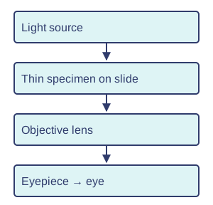
Microscopy — Place a thin specimen on a slide, add a suitable stain if instructed, and lower the coverslip carefully to reduce bubbles. Start with low power, focus safely and then increase magnification. Draw clear outlines, label structures and include a scale or magnification.
Microscope light path — Light passes through a thin specimen before reaching the objective and eyepiece. A thick opaque specimen blocks the light and hides internal detail.Light through a microscope. Original schematic; see the explanation for its limits. Full-size diagram
Focusing safely — Locate the specimen at low magnification first. Use the focusing procedure taught for that microscope; avoid driving the objective into the slide. Higher magnification narrows the field of view.
Biological drawing — Use clear single outlines and straight label lines that end at the named structures. Draw what can actually be observed; shading or invented organelles does not improve accuracy.
Magnification — Total microscope magnification is eyepiece magnification × objective magnification: ×10 and ×20 give ×200. For a drawing, magnification = image size ÷ actual size, using the same units. A 20 mm image of a 0.1 mm structure is ×200; magnification has no unit.
Unit conversion for images — Convert measurements to the same unit before dividing: 1 mm = 1000 micrometres. An image 30 mm wide of a cell 0.15 mm wide has magnification 30 ÷ 0.15 = ×200.
Magnification and resolution — Magnification makes an image larger. Resolution concerns distinguishing nearby features as separate. Enlarging a blurred image does not reveal new detail.
Structure and function
Specialisation — A cell's shape and structures support its job. A root hair cell has a long extension for absorbing water and minerals; a nerve cell carries signals over distances; a red blood cell transports oxygen. These examples illustrate structure linked to function.
Root hair adaptation — A root hair's long extension increases contact area with soil water. A thin boundary gives a short exchange distance; the cell's shape supports absorption rather than pulling whole soil particles inside.Specialised shapes support different functions. Original schematic; see the explanation for its limits. Full-size diagram
Red blood cells — Human red blood cells contain haemoglobin that carries oxygen. Their biconcave shape, like a disc with a dip on both faces, gives a large surface area relative to volume. This provides more surface for oxygen exchange compared with the amount of cell material; mature cells lack a nucleus, leaving more space for haemoglobin.
Nerve and muscle cells — A nerve cell's long extension helps carry electrical signals between distant parts. Muscle cells contain structures that shorten and use energy during contraction. Specialised shape and contents support different jobs.
Diffusion — Concentration describes how much of a substance there is in a given volume. Diffusion is the net movement of particles from higher to lower concentration through random movement. Net means the overall change after counting movement in both directions. Oxygen diffuses into cells and carbon dioxide diffuses out when suitable concentration differences exist. Particles still move in both directions.
Diffusion after mixing — Particles continue moving when concentrations become equal. Equal average movements in opposite directions give no net transfer, rather than all particles stopping.Diffusion and dynamic equilibrium. Original schematic; see the explanation for its limits. Full-size diagram
Diffusion rate — A bigger concentration difference usually increases net diffusion. A larger exchange area and shorter distance also help exchange, explaining why thin, extensive surfaces are useful in organs.
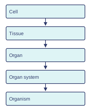
Organisation — In a multicellular organism, similar cells form tissues; several tissues form an organ; organs cooperate in an organ system. Muscle tissue, the heart and the circulatory system illustrate these levels. The whole organism depends on its systems working together.Levels of organisation. Original schematic; see the explanation for its limits. Full-size diagram
From cells to systems — Cardiac muscle cells form muscle tissue in the heart. The heart contains several tissues and acts as an organ within the circulatory system. A tissue and an organ are different levels of organisation.
Unicellular life — Amoeba can take in food and respond to its surroundings as a single cell. Its flexible shape and extensions help engulf food; a unicellular alga has chloroplasts suited to photosynthesis. Some unicellular organisms photosynthesise. Their one cell must exchange materials, release energy and reproduce; they do not have multicellular organs.
Unicellular organisms — One amoeba cell takes in food, exchanges gases and responds to its surroundings. It does not have a digestive system with a stomach and intestines; one cell performs the necessary processes.
Dependence — Digestive organs supply absorbed nutrients; gas exchange supplies oxygen; transport moves substances; cells respire. A change in one system can affect others. Function cannot be explained by looking at a single organ in isolation.
Systems cooperate — After digestion, soluble nutrients enter the blood. Gas exchange adds oxygen; circulation delivers both to cells, where respiration releases usable energy. These processes occur in different places but depend on one another.
Test yourself
30 questions · Random sets of 10. These quick checks support revision; practise longer explanations and justified judgements too.
Mind map
Use the branches to recall the ideas and explain their connections. Check the revision notes for the full detail.
KS3SCI2 · Cells 1 / Cells 2 / Cells 3 / Cells 4
View KS3SCI2 · Cells 1 / Cells 2 / Cells 3 / Cells 4 mind mapOpen the full-size map to zoom. Use the printable view for A4 landscape or enlarge to A3.
View KS3SCI2 · Cells 5 / Cells 6 / Functions 1 / Functions 2 mind mapOpen the full-size map to zoom. Use the printable view for A4 landscape or enlarge to A3.
View KS3SCI2 · Functions 3 / Functions 4 / Functions 5 mind mapOpen the full-size map to zoom. Use the printable view for A4 landscape or enlarge to A3.
 Mr Kidman's Classroom