Trace a stimulus-response pathway — A stimulus is a change in internal or external conditions. A receptor detects a relevant change, coordination links that information to appropriate activity and an effector, such as a muscle or gland, produces a response. The response can improve survival or reproduction in context; sensing a stimulus does not imply conscious awareness or a response that is always beneficial.
Explain growth factors and IAA — Indoleacetic acid, or IAA, is an auxin growth regulator produced in growing regions and transported to responsive tissues. Its effect on cell elongation depends on concentration and tissue sensitivity. Roots and shoots can respond differently to the same concentration, so avoid a universal statement that IAA always increases growth in every plant tissue.
Explain shoot phototropism — In the standard young-shoot model, light from one side (unilateral light) causes an unequal IAA distribution, with greater elongation on the shaded side under suitable conditions. Unequal extension bends the shoot towards light. The shoot does not move because its cells migrate towards the lamp, and the illuminated side is not necessarily the side that lengthens fastest.
Explain gravitropism using tissue sensitivity — In a horizontal shoot, redistribution of IAA can increase elongation on the lower side so the shoot curves upwards. In a root, a higher concentration on the lower side can inhibit elongation relative to the upper side, producing downward curvature. Relate direction to the stated dose-response relationship rather than assuming roots and shoots have identical sensitivity.
Evaluate growth investigations — Compare seedlings of similar age and size under controlled light, temperature, water and exposure conditions, using a suitable concentration range and controls. Measure change in length or curvature consistently with repeats. Removing or applying material at a tip can affect more than IAA alone, so interpret experimental evidence using its actual controls.
Distinguish taxis and kinesis — A taxis is directional movement towards or away from a stimulus, while a kinesis changes movement rate or turning frequency without a fixed direction relative to the stimulus. For example, moving more rapidly and turning less in unfavourably dry conditions can increase the chance of leaving them without directly navigating towards a humidity source.
Plan a choice-chamber or maze investigation — Required practical 10 investigates how an environmental variable affects animal movement. Define a measurable outcome, offer comparable accessible regions, vary the named factor and control temperature, light, time and starting position where appropriate. Rotate or randomise apparatus orientation and use repeats to distinguish a treatment preference from a fixed side bias.
Evaluate behavioural evidence and welfare — Numbers found in a chamber at one time show distribution, not necessarily the exact movement rule producing it. Observe paths or movement rates when distinguishing taxis from kinesis. Use suitable animals, humane conditions and an approved protocol, avoid harmful extremes and minimise disturbance; repeated readings from the same individuals are not automatically independent animals.
Explain a three-neurone reflex — A sensory neurone carries information from a receptor to a relay neurone, which communicates with a motor neurone leading to an effector. A rapid protective response can occur before conscious interpretation of the event, although information can also reach the brain. Synapses provide sites for direction and integration; a reflex is not evidence that every response bypasses all coordination.
Receptors, rods and cones
Explain a Pacinian corpuscle — A Pacinian corpuscle contains a sensory ending surrounded by layers that transmit suitable deformation. Mechanical pressure or vibration changes the shape of stretch-mediated sodium channels in the ending. Increased sodium entry changes membrane potential, providing a generator potential rather than directly making an antibody or a hormone.
Distinguish generator and action potentials — A generator potential is graded: stronger suitable stimulation can produce a greater change over the responsive range. If the trigger region reaches threshold, action potentials arise in the sensory neurone. Stronger stimulation is commonly represented by a higher impulse frequency, not by each action potential becoming proportionally larger.
Relate rods to low-light sensitivity — Rod photoreceptors have pigments suited to dim-light detection. Convergence, where signals from several rods feed into a shared pathway, can allow small contributions to add together (summate), improving sensitivity while reducing precise localisation. Rod-based vision has limited colour discrimination; high sensitivity is therefore associated with different performance from fine colour vision.
Relate cones to colour and acuity — Cone types have different spectral sensitivities, enabling colour discrimination through comparison of their activity. Less convergence in suitable retinal regions supports more precise localisation and high visual acuity, meaning the ability to distinguish fine detail, but stronger illumination is needed than for rod-mediated sensitivity. Different cone responses are not separate coloured pictures sent from each receptor.
Compare retinal distributions — The foveal region is specialised for high-acuity cone-mediated vision, while rods are abundant in other suitable retinal regions. At the optic disc, fibres leave the eye and photoreceptors are absent, producing a blind spot. Interpret a provided distribution graph without assuming all regions contain the same receptor mix.
Myogenic rhythm and heart-rate control
Trace the heart's electrical sequence — The sinoatrial node initiates electrical activity in the myogenic heart, spreading through atrial tissue and promoting atrial contraction. Activity reaches the atrioventricular node, where a delay allows filling before ventricular activation. The pathway proceeds through the bundle of His and Purkyne tissue, coordinating ventricular contraction from the lower regions upwards.
Explain myogenic rather than nerve-initiated contraction — Cardiac tissue can initiate rhythmic activity without each beat requiring a motor impulse from outside the heart. Autonomic nerves alter the intrinsic rhythm to match conditions. The nervous system therefore regulates heart rate rather than serving as the sole source of every normal heartbeat.
Use chemoreceptor feedback — Chemoreceptors in appropriate regions, including major arterial sites and central monitoring pathways, provide information related to carbon dioxide, pH or oxygen as relevant to the receptor. Increased carbon dioxide and associated pH change can lead the cardiovascular control centre to increase sympathetic influence on the SAN. Describe the supplied signal and pathway rather than treating every receptor as measuring identical variables.
Use pressure-receptor feedback — Baroreceptors in regions such as the carotid sinus and aortic arch respond to arterial stretch. Increased pressure can increase their activity and promote responses reducing heart rate through autonomic control, while reduced pressure can produce the opposite adjustment. Pressure is sensed through mechanical stretch, not by counting the number of glucose molecules in the blood.
Compare autonomic effects — The autonomic nervous system controls activities such as heart rate without conscious direction. Its sympathetic and parasympathetic divisions can have opposing effects. Sympathetic input generally increases SAN rate, while parasympathetic vagal input generally slows it through different chemical signalling at the target. Heart-rate change can alter cardiac output, but output also depends on stroke volume. Evaluate a response using both rate and volume information when provided.
Resting potentials and action potentials
Relate motor-neurone structure to function — A motor neurone has a cell body, input regions, a long axon and terminal branches communicating with target cells. In a myelinated axon, insulating sheath segments are separated by nodes of Ranvier. Signals travel as changing membrane potentials, not as a continuous stream of neurotransmitter flowing the entire axon length.
Explain the resting ion distribution — The sodium-potassium pump uses ATP to move three sodium ions out for two potassium ions in, helping maintain unequal ion distributions. Differential membrane permeability, especially potassium leak at rest, and electrochemical gradients contribute to a negative internal potential. The pump maintains gradients; it is not the only cause of the instantaneous membrane voltage.
Trace depolarisation — If adequate local stimulation reaches threshold, voltage-gated sodium channels open, increasing sodium permeability. Sodium enters down its electrochemical gradient, rapidly making the membrane potential less negative and then positive in the standard action-potential model. Opening a channel changes permeability; the channel does not create sodium ions.
Trace repolarisation and recovery — Sodium channels become inactivated and voltage-gated potassium channels open with a delay, allowing potassium to leave. The potential returns towards and can briefly pass below resting level before channels reset. Immediate repolarisation is mainly explained by changing channel permeability, not by the sodium-potassium pump suddenly reversing its direction.
Explain all-or-nothing signalling — Once threshold is reached, a normal action potential has a characteristic amplitude in that neurone. A weaker subthreshold input does not produce a proportionally miniature action potential. Stimulus strength can be encoded by impulse frequency and recruitment of neurones, while generator potentials and postsynaptic potentials remain graded.
Explain propagation in an unmyelinated axon — Local currents from an active region depolarise an adjacent region to threshold, triggering a new action potential there. The wave is regenerated along the membrane rather than one identical sodium ion travelling all the way to the terminal. Refractory membrane behind the advancing wave helps prevent immediate backward re-excitation.
Explain the refractory period — During the absolute refractory interval, channels are not ready to support another normal action potential, regardless of an ordinary additional stimulus. Later recovery can require a stronger stimulus. Refractoriness separates impulses, limits maximum frequency and supports one-way propagation along a stimulated axon in the usual physiological sequence.
Calculate a frequency limit — If the relevant minimum interval between impulses is 2.5 ms, convert it to 0.0025 s and calculate 1/0.0025 = 400 s⁻¹, or 400 Hz. This is a theoretical maximum under the stated assumption, not a guarantee that the neurone normally fires at that rate.
Explain saltatory conduction — Myelin reduces ion leakage and the need for membrane depolarisation along insulated segments; action potentials are regenerated at nodes of Ranvier. Local currents spread between nodes, making conduction faster than otherwise comparable unmyelinated pathways. Saying an impulse jumps is shorthand, not evidence that electrical influence crosses an empty break without a physical pathway.
Compare other conduction factors — A larger axon diameter reduces resistance to internal local-current spread and can increase conduction speed. Moderate temperature increases can speed channel processes, while excessive heat can impair function. Compare like conditions and distinguish conduction velocity from impulse frequency: a faster travelling impulse does not automatically have a larger amplitude.
Cholinergic synapses and integration
Relate synaptic structures to roles — Presynaptic means on the sending side of a synapse; postsynaptic means on the receiving side. A presynaptic terminal contains neurotransmitter vesicles and voltage-gated calcium channels. A narrow synaptic cleft separates it from the postsynaptic membrane carrying suitable receptors. The direction of chemical transmission reflects transmitter release and receptor placement rather than a freely bidirectional electrical wire across the gap.
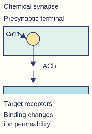
Trace transmitter release — An arriving action potential depolarises the presynaptic terminal and opens calcium channels. Calcium entry stimulates vesicle fusion with the membrane and release of acetylcholine by exocytosis. Acetylcholine diffuses across the cleft; the action potential itself does not leap into the next cell as a packet of chemical.Calcium entry in the sending terminal triggers acetylcholine release. Acetylcholine crosses the cleft and binds receptors on the receiving membrane; receptor channels, breakdown and recycling are described in the adjoining notes.
Trace the postsynaptic response — Acetylcholine binds complementary receptors and opens appropriate ligand-gated ion pathways, producing a postsynaptic potential. At an excitatory cholinergic synapse this can depolarise the membrane towards threshold. Whether an action potential follows depends on integrated input, so one small excitatory event need not always trigger an impulse.
End the acetylcholine signal — Acetylcholinesterase hydrolyses acetylcholine, limiting continued receptor stimulation. Breakdown products such as choline can be taken up and reused in transmitter synthesis, which requires metabolic support. Ending the signal allows discrete responses; otherwise persistent receptor activation can disrupt normal signalling rather than simply improve it.
Explain temporal and spatial summation — Temporal summation combines postsynaptic effects arriving close together in time from repeated activity at a synapse. Spatial summation combines effects from different synapses. Inputs must overlap sufficiently in their effects to help reach threshold; summation concerns graded local potentials, not adding two action-potential amplitudes into one giant impulse.
Explain inhibition — An inhibitory synaptic input can make reaching threshold less likely by moving membrane potential away from threshold or increasing conductance that counteracts excitation. Depending on the system, ion pathways involving chloride or potassium can contribute. Use the provided mechanism rather than assuming every neurotransmitter produces excitation at every receptor.
Predict drug effects from mechanisms — A receptor agonist may mimic transmitter action, an antagonist may block it and an enzyme inhibitor may prolong transmitter availability. A release inhibitor can reduce signalling even if receptors remain intact. Follow the named target through release, binding, ion movement and signal termination, and distinguish an initial effect from prolonged dysfunction.
Skeletal muscle and sliding filaments
Explain antagonistic skeletal action — Skeletal muscles generate tension by contracting and pull on attachment points; they do not actively push an opposing bone away. Antagonistic muscles allow movement in opposite directions around a joint, supported by a relatively incompressible skeleton. Relaxation permits lengthening under other forces rather than actively driving the reverse movement by itself.
Relate muscle hierarchy to structure — A skeletal muscle contains bundles of fibres; each fibre contains many myofibrils composed of repeating sarcomeres. Thick filaments contain myosin and thin filaments contain actin with associated regulatory proteins. Sarcomere boundaries are Z lines, and regular filament arrangement produces the striated, or striped, appearance.
Interpret sarcomere changes — During contraction, thin and thick filaments slide relative to each other without each filament becoming shorter. Z lines move closer, the I band and H zone decrease, and the A band corresponding to thick-filament length remains approximately constant. Distinguish shortening of the sarcomere from shortening of an individual actin or myosin filament.Simplified paired sarcomeres: boundary lines move closer while thin filaments (teal) and thick filaments (orange) retain their lengths. Increased overlap produces shortening; detailed bands and multiple filament rows are omitted.
Link neuromuscular transmission to calcium release — At a neuromuscular junction, motor-neurone acetylcholine release produces an end-plate response that normally triggers a muscle action potential. Electrical activity spreads over the fibre and into its internal membrane system, leading to calcium release from the sarcoplasmic reticulum, a specialised internal membrane network that stores calcium in muscle fibres. This calcium release is inside the muscle and should not be confused with presynaptic calcium entry causing transmitter release.
Expose binding sites — In resting muscle, regulatory protein positioning limits myosin binding to actin. Increased calcium changes the regulatory complex so tropomyosin moves and exposes binding sites. The resulting access permits cross-bridge formation; calcium does not itself hydrolyse all the ATP or shorten the actin molecules.
Trace the cross-bridge cycle — An energised myosin head binds exposed actin, then changes orientation during the power stroke as products are released. ATP binding causes detachment from actin, and ATP hydrolysis re-energises the head for another cycle. Repeat cycles slide filaments while suitable calcium and ATP conditions persist; ATP is needed for detachment as well as supporting continued activity.
Explain relaxation and phosphocreatine — Calcium is actively returned to storage, allowing regulatory proteins to cover binding sites and reducing cross-bridge cycling. Phosphocreatine can transfer phosphate to ADP to replenish ATP rapidly during short high-demand activity. It is a finite immediate buffer, not a permanent substitute for respiration or a molecule that directly pulls actin.
Compare slow and fast fibres — Slow fibres generally have abundant mitochondria, myoglobin and capillary supply and resist fatigue during sustained aerobic work. Fast fibres support rapid powerful activity, often with greater reliance on rapid ATP supply and glycolytic capacity, and can fatigue sooner. Real fibre categories vary; apply the supplied structural evidence rather than assuming all fast fibres have no mitochondria.
Feedback and blood-glucose regulation
Define homeostasis precisely — Homeostasis maintains the internal environment within limits through regulated physiological responses, not at one perfectly fixed value with no fluctuation. Temperature and pH affect enzyme activity, while glucose provides respiratory substrate and influences blood water potential. A control system needs information about the relevant variable and effectors capable of changing it.
Explain negative and positive feedback — Negative feedback produces responses opposing a departure from an appropriate range; as the departure diminishes, the corrective response is reduced. Separate mechanisms can correct deviations in opposite directions. Positive feedback amplifies an initial change until an endpoint or external limit intervenes, so it should not be described as the routine stabilising mechanism in every homeostatic loop.
Distinguish liver glucose pathways — Glycogenesis stores glucose as glycogen, glycogenolysis breaks glycogen down and gluconeogenesis forms glucose from suitable noncarbohydrate precursors such as glycerol and some amino acids. The similar names describe different directions and substrates. Liver glucose output helps regulate the concentration available to other tissues.
Trace insulin signalling — Raised blood glucose stimulates pancreatic beta cells to release insulin. Binding to suitable target receptors can increase uptake through regulated insertion of glucose transporters in responsive tissues and promote glycogen synthesis. These transporters are carrier proteins rather than unrestricted aqueous pores, and not every tissue's glucose uptake is insulin dependent.
Trace glucagon signalling — When blood glucose falls, pancreatic alpha cells release glucagon, which acts on suitable liver receptors to promote glycogen breakdown and glucose formation from appropriate precursors. Glucose can then be released into blood. Glucagon does not simply push glycogen out of liver cells as intact granules.
Explain the second-messenger pathway — For the relevant glucagon or adrenaline pathway, hormone binding at a surface receptor activates a pathway involving adenylate cyclase, which generates cAMP from ATP. Cyclic AMP (cAMP) acts as a second messenger inside the cell, carrying the signal from the surface receptor. It activates protein kinase, an enzyme that transfers phosphate to proteins, starting a phosphorylation cascade in which successive enzyme activations affect target enzymes. Amplification allows a small extracellular signal to alter many intracellular molecules without the hormone entering to become the enzyme itself.
Relate adrenaline to glucose availability — Adrenaline can stimulate liver glycogen breakdown through appropriate receptor and second-messenger pathways, increasing glucose availability during acute demand. It also has other effects that depend on tissue and receptor type. Do not infer that all hormones use exactly the same receptors or produce the same response in every target cell.
Compare types of diabetes — Type 1 diabetes involves loss of effective insulin production, commonly through autoimmune destruction of beta cells. Type 2 involves reduced tissue responsiveness to insulin with varying impairment of secretion. Their management can include different combinations of insulin, other prescribed treatments and dietary measures; these mechanisms should not be collapsed into the claim that everyone with diabetes produces no insulin.
Evaluate population evidence responsibly — Associations of type 2 diabetes incidence with diet, body composition, activity and social conditions require attention to confounding, access and variation between individuals. Evaluate positions of health advisers and food producers using study quality, outcomes and possible conflicts of interest. Avoid implying that one exposure alone explains every case or that a population association establishes an individual's cause.
Glucose calibration practical
Prepare glucose standards — Required practical 11 uses a dilution series and colorimetry to estimate glucose in an unknown simulated urine sample. Use known stock concentration and measured volumes, with C₁V₁ = C₂V₂ when applicable. To make 10 cm³ of 2 g dm⁻³ from a 10 g dm⁻³ stock, use 2 cm³ stock and dilute to 10 cm³ total, not add 10 cm³ water.
Use a consistent assay — Apply the approved glucose assay with equal reagent volumes, reaction time and temperature for standards and unknowns. Select a suitable wavelength and blank, and ensure measurement is appropriate to the preparation rather than reading an unsettled precipitate as if it were a clear solution. A curve is valid only for the method and working range used.
Interpret the calibration curve — Plot instrument response against known concentration, use a suitable fitted relationship and interpolate the unknown within the calibrated range. If the unknown was diluted fivefold and the assayed solution is estimated at 0.8 g dm⁻³, the original concentration is 4.0 g dm⁻³. Above-range measurements require appropriate dilution rather than confident extrapolation far beyond the standards.
Evaluate precision and interpretation — Replicate preparation and measurement, include appropriate controls and consider pipetting and instrument uncertainty. An educational simulated-sample assay demonstrates measurement skills, not a complete clinical diagnosis. Distinguish a concentration estimate from evidence about why glucose is present or how a particular person should be treated.
Nephrons and selective reabsorption
Trace the nephron pathway — Blood enters glomerular capillaries within Bowman's capsule, and filtrate proceeds through the proximal convoluted tubule, loop of Henle, distal convoluted tubule and collecting duct. Blood remaining in vessels follows associated capillary pathways. The tubule contains filtrate while surrounding vessels contain blood; do not merge them into a single open tube.
Explain ultrafiltration — Relatively high glomerular hydrostatic pressure drives water and small dissolved substances across a selective filtration barrier into the capsule. Blood cells and most large plasma proteins are retained. Filtration is not the selective decision to remove only waste: useful small solutes such as glucose also enter the filtrate and must be recovered.
Explain proximal glucose reabsorption — In the proximal tubule, suitable sodium-linked transport brings glucose into epithelial cells using gradients maintained by ATP-dependent sodium pumping. Glucose exits towards surrounding blood through appropriate transporters. Large membrane area and many mitochondria support reabsorption; transport capacity can be exceeded when filtered glucose loads are sufficiently high.
Explain proximal water reabsorption — Reabsorption of solutes alters water-potential relationships so water follows across suitable pathways by osmosis. Much filtered water is recovered before the collecting duct, while later regulated reabsorption adjusts final output. Water is not hydrolysed into ATP by the tubule cells to remove it from filtrate.
Explain the loop of Henle gradient — In the standard model, the descending limb is relatively permeable to water and loses water into the increasingly concentrated medulla, the kidney's inner region. The ascending limb limits water passage while sodium chloride moves out, including active transport in the thick region. Opposite flow directions and differing permeability help maintain a medullary gradient useful for later water recovery.
Connect the gradient to concentration — Fluid leaving the ascending region can be relatively dilute, while the medullary interstitial environment is concentrated. The collecting duct passes through this gradient, allowing water removal when its permeability is increased. The loop's function is not that both limbs freely lose identical amounts of water and sodium at every level.
ADH and blood-water-potential control
Detect reduced blood water potential — Osmoreceptive cells in the hypothalamus respond to changes associated with blood water potential. When water potential falls, increased ADH release supports greater water recovery. ADH is synthesised in hypothalamic neurones and released from their terminals in the posterior pituitary, rather than being made by kidney collecting ducts.
Trace ADH action — ADH binds receptors on suitable distal nephron and collecting-duct cells, triggering signalling that increases membrane insertion of aquaporin water channels. The cells become more water permeable, allowing greater osmosis into the lower-water-potential medulla and onwards to blood. This produces a smaller volume of more concentrated urine when other conditions support the gradient.
Explain the opposite response — After sufficient water intake, blood water potential rises towards the regulated range, reducing the stimulus for ADH release. Lower collecting-duct water permeability allows more water to remain in tubular fluid, giving a larger volume of more dilute urine. Negative feedback limits the corrective response as the monitored departure is reduced.
Interpret water-balance evidence — Use urine volume, concentration, water intake and hormone information together. A small urine volume can reflect several influences, and concentration is a ratio rather than the total amount of every solute excreted. Predict a mechanistic response from the stated conditions without assuming one measurement alone identifies every cause.
Test yourself
50 questions · Random sets of 10. These quick checks support revision; practise longer explanations and justified judgements too.
 Mr Kidman's Classroom