Shared biochemical principles — Living organisms use the same broad groups of carbon-based molecules and many similar biochemical reactions. This shared chemistry is consistent with common ancestry, but similarity of one molecule alone does not establish a complete evolutionary relationship. Connect molecular structure to what a cell needs to do rather than learning a list of substances without functions.
Monomers and polymers — A monomer is a smaller molecular unit that can join with others to form a larger molecule. A polymer contains many linked monomers: amino acids form polypeptides, nucleotides form polynucleotides and monosaccharides form polysaccharides. Not every large biological molecule is a polymer; a triglyceride does not have a chain of repeating monomer units.
Condensation forms a linkage — A condensation reaction joins molecules through a chemical bond while eliminating water. Joining two monosaccharides forms a glycosidic bond; amino acids form peptide bonds; nucleotides form phosphodiester links. Identify the reacting groups and the bond in a diagram rather than assuming that water simply sticks the molecules together.
Hydrolysis uses water to break a linkage — Hydrolysis breaks a chemical bond through reaction with water. Parts of the water molecule become attached to the separated products. Digestion uses enzyme-catalysed hydrolysis to produce smaller absorbable molecules; it does not merely dissolve a polymer or turn every food molecule into glucose.
Count bonds and water molecules — Joining five monomers into one unbranched chain requires four links and eliminates four water molecules. Hydrolysing those four links uses four water molecules. State what is being counted: this relationship applies to a chain constructed from separate monomers and does not justify treating every complex branched or cyclic molecule as an identical structure.
Carbohydrate structure and function
Recognise monosaccharides — Glucose, galactose and fructose are monosaccharides. Glucose is a hexose, a six-carbon monosaccharide, with the formula C6H12O6, but a formula alone does not tell you how atoms are arranged. Structural differences between molecules with the same molecular formula help explain why enzymes recognise particular substrates rather than every sugar indiscriminately.
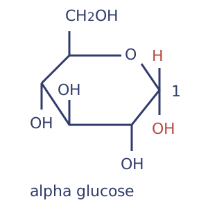
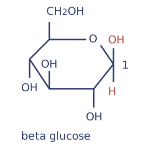
Distinguish alpha and beta glucose — In the conventional glucose ring diagrams used here, alpha glucose has its carbon-1 hydroxyl group below the ring and beta glucose has it above, relative to the standard orientation. The difference concerns the arrangement around carbon 1, not a different molecular formula. Reorienting an entire drawing does not turn one isomer into the other.Alpha glucose: carbon-1 hydroxyl below the ring in this fixed orientation. Full-size diagramBeta glucose: carbon-1 hydroxyl above the ring in the matching orientation. Full-size diagram
Form disaccharides correctly — Maltose is formed from two glucose molecules; sucrose from glucose and fructose; lactose from glucose and galactose. Condensation creates a glycosidic bond and releases water. For two hexoses, C6H12O6 + C6H12O6 gives C12H22O11 + H2O; simply adding the two starting formulae misses the eliminated water.
Amylose supports compact storage — Amylose is a component of starch made from alpha glucose joined predominantly through alpha-1,4 glycosidic bonds. Its chain coils, allowing glucose units to be stored compactly. It is a storage material rather than a membrane-crossing supply of individual glucose molecules; hydrolysis is needed to release smaller sugars.
Amylopectin adds branching — Amylopectin contains alpha-1,4 links along chains and alpha-1,6 links at branch points. Branching provides more ends where enzymes can act, allowing stored glucose to be released quickly when suitable enzymes are present. This release from a store is called mobilisation. Distinguish the branching of the polymer from the coiling of an individual chain.
Starch and osmotic effects — Starch is an insoluble glucose-storage mixture in plants. Storing many glucose units as a polymer reduces the osmotic effect compared with keeping the same number as freely dissolved glucose molecules. There are fewer separate dissolved particles to lower water potential, so the store has less tendency to draw water into the cell by osmosis. Insolubility also helps retain the store inside cells; the cell still needs enzymes to mobilise it when required.
Glycogen is a branched store — Glycogen is a highly branched alpha-glucose polymer stored by animals and other organisms including fungi. Numerous ends make glucose mobilisation possible at many sites. Do not confuse glycogen, the polymer store, with glucagon, the hormone involved in regulating blood glucose.
Cellulose forms straight chains — Cellulose contains beta-glucose units joined by beta-1,4 links, with alternate glucose units rotated in relation to one another. This arrangement gives straight chains rather than the coiled arrangement associated with amylose. The same glucose formula therefore supports very different properties through a different linkage and arrangement.
Microfibrils resist tensile forces — Hydrogen bonds between neighbouring cellulose chains help hold them together in microfibrils. A microfibril is a bundle of these parallel chains. Together, the many hydrogen bonds provide tensile strength, meaning resistance to being pulled apart, in plant cell walls and help resist expansion when water enters. The glycosidic links within a chain are covalent; the hydrogen bonds joining chains are a different type of interaction.
Explain specificity in digestion — Human digestive enzymes can hydrolyse particular carbohydrate linkages, but humans do not make cellulase to digest cellulose. A statement that every polysaccharide becomes glucose during human digestion is therefore incorrect. The structure of the substrate and the active site's specificity determine whether a particular enzyme can catalyse its hydrolysis.
Lipids and membrane-forming properties
Build a triglyceride — A triglyceride forms from one glycerol and three fatty acids. Glycerol has three hydroxyl groups, each of which can react with a fatty acid's carboxyl group. Three condensation reactions produce three ester bonds and three water molecules. Lipid is a broad category; triglycerides are not long chains of repeating identical monomers.
Recognise fatty acids — A fatty acid contains a carboxyl group (–COOH) and a hydrocarbon region made mainly of carbon and hydrogen, often represented together as RCOOH. Chain length and carbon-carbon bonding affect packing and properties. In a biological diagram, identify the carboxyl end and hydrocarbon portion rather than treating the letter R as a particular named element.
Compare saturation — A saturated fatty acid has no carbon-carbon double bonds in its hydrocarbon chain; an unsaturated one has one or more. A cis double bond introduces a bend that can reduce close packing. Not every unsaturated arrangement has the same geometry, so relate a structural diagram to packing rather than assuming the word unsaturated alone specifies every property.
Relate packing to physical properties — Fatty-acid composition influences how closely lipid molecules pack and therefore affects melting behaviour. Mixtures also depend on chain length and proportions, so a comparison needs the stated structures or data. Avoid claiming that every lipid of animal origin must be solid or that every plant lipid must be liquid.
Explain hydrophobic storage — Triglycerides are largely non-polar: their molecules have little separation into positively and negatively charged regions. They do not mix readily with water, so are described as hydrophobic. They provide a compact energy store with little associated water, and their oxidation can release a large amount of energy per gram. This is a comparison of overall respiratory reactions, not a claim that breaking an isolated bond releases energy without any other changes.
Describe a phospholipid — A typical membrane phospholipid has a glycerol-based structure with two fatty-acid tails and a phosphate-containing polar head. The head can interact with water while the tails are hydrophobic. Use the diagram's actual groups when comparing it with a triglyceride; a phospholipid is not simply a triglyceride with an extra fourth fatty acid.
Explain bilayer formation — In an aqueous environment, phospholipids can arrange with hydrophilic (water-attracting) heads facing water and hydrophobic tails, which do not interact readily with water, facing away from it. In a bilayer, the tails face an internal hydrophobic region between two head layers. These properties help form the structural basis of cell membranes, while membrane proteins provide additional functions considered in Cells.
Proteins, bonding and molecular shape
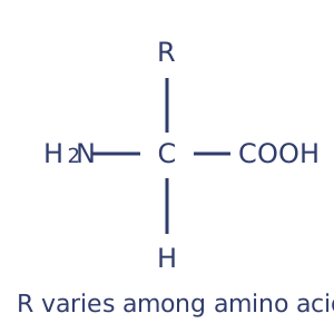
Recognise an amino acid — An amino acid has an amino group, a carboxyl group, hydrogen and a variable R group attached to a central carbon. The 20 common amino acids differ in their side chains. The neutral diagram is a useful structural representation; ionisation in solution depends on conditions, so it should not be taken as proof that every amino acid has no charge at every pH.General amino-acid shorthand; ionic form depends on conditions. Full-size diagram
Form peptide bonds — Condensation between the amino group of one amino acid and the carboxyl group of another forms a peptide bond and releases water. Two joined amino acids form a dipeptide; many form a polypeptide. A functional protein may contain one polypeptide or several associated chains, so polypeptide and functional protein are not always interchangeable terms.
Primary structure determines possibilities for folding — Primary structure is the sequence of amino acids in a polypeptide. It determines the positions and chemical properties of the side chains that can interact during folding. Changing one amino acid may affect function greatly, slightly or not detectably, depending on its position and role; it does not automatically destroy every protein.
Secondary structure uses backbone interactions — Alpha helices and beta-pleated sheets are examples of secondary structure. Hydrogen bonds involving parts of the polypeptide backbone stabilise these patterns. Do not explain every level of protein structure as bonding between R groups: side-chain interactions are particularly important when considering tertiary folding.
Tertiary structure creates a specific overall fold — Tertiary structure is the overall three-dimensional folding of one polypeptide. Hydrogen bonds, ionic interactions, disulfide bridges and hydrophobic interactions can contribute. The resulting shape and distribution of chemical groups affect binding and solubility; listing bond names without explaining their role does not establish a structure-function relationship.
Disulfide bridges differ from weaker interactions — A disulfide bridge is a covalent link formed between suitable cysteine side chains. Ionic interactions depend on charges. Hydrogen bonds are a different type of attraction and can occur between molecules (intermolecular) or within one molecule (intramolecular). Changes in pH can alter charges and interactions; do not assume that every change in folding necessarily breaks the peptide bonds of the primary sequence.
Quaternary structure involves associated chains — Quaternary structure describes the arrangement of multiple polypeptide chains in a functional protein. Haemoglobin is an example with four polypeptide chains and haem groups, whereas a protein with only one polypeptide has no multi-chain quaternary arrangement. Additional non-protein groups may be important to function but are not themselves polypeptide chains.
Connect protein properties to function — Fibrous arrangements such as collagen support tensile strength; globular proteins can expose particular binding surfaces or active sites. A structure-function answer should identify the relevant feature and explain its consequence. Do not claim that all proteins are enzymes or that all proteins have identical solubility and mechanical properties.
Explain denaturation carefully — Temperature or pH changes can disrupt interactions maintaining a protein's functional shape. Denaturation can alter binding sites and activity without hydrolysing the primary peptide sequence under ordinary experimental conditions. An enzyme is not a living organism and is not killed; describe the structural change and its functional consequence.
Enzyme action and factors affecting rate
Enzymes lower activation energy — An enzyme provides a reaction pathway with a lower activation energy, the energy barrier that reactants must overcome. More reactant molecules can therefore react per second, enabling a faster reaction under suitable conditions. It is not used up overall and does not supply the reaction's substrate. A catalyst changes the rate, not the overall energy difference between the same reactants and products.Qualitative paths share starting and finishing levels; enzyme catalysis lowers the barrier. Full-size diagram
Induced fit develops the active interaction — A substrate interacts with an active site whose shape and chemical groups are suitable for binding. Binding can induce a change in the enzyme's conformation, or three-dimensional shape, that improves the catalytic interaction. This explains more than a completely rigid lock-and-key picture: the enzyme is dynamic rather than a solid mould.
Specificity depends on structure and chemistry — The active site's tertiary structure places particular groups in positions that interact with a suitable substrate. A different shape or chemical arrangement may prevent an effective enzyme-substrate complex. Specificity concerns productive binding and catalysis, not merely whether a molecule can collide with the enzyme.
Temperature has competing effects — At lower temperatures, increasing temperature generally increases kinetic energy and the frequency of productive collisions. At sufficiently high temperatures, disruption of the enzyme's structure reduces the number of functional active sites. The observed optimum depends on the enzyme and conditions, including the duration of exposure; it is not universally 37 degrees Celsius.
Low temperature is not ordinary thermal denaturation — Cooling commonly reduces activity because molecules move more slowly, while warming can restore a higher rate if the enzyme remains structurally functional. Compare this reversible kinetic effect with lasting loss of active-site shape after excessive heating. Do not infer denaturation simply because a cold sample reacted slowly.
pH changes ionisation and interactions — Changing pH alters hydrogen-ion concentration and may change charges on groups involved in binding or maintaining structure. Activity may fall as productive binding becomes less likely, and extreme conditions can disrupt the functional fold. Different enzymes have different pH optima; neutral pH is not automatically best for every enzyme.
Substrate concentration can become limiting — At low substrate concentration, adding substrate increases opportunities to form enzyme-substrate complexes. With a fixed enzyme concentration, rate eventually approaches a plateau when active sites are operating at their available capacity under those conditions. A plateau does not mean that the enzyme has been used up.
Enzyme concentration matters when substrate is available — Increasing enzyme concentration provides more active sites and can increase rate if sufficient substrate is present and other conditions do not become limiting. If the substrate is scarce, adding more enzyme may have little effect. Interpret a graph using its controlled conditions rather than assuming proportionality at every concentration.
Competitive inhibition involves competing binding — In the simple competitive model, an inhibitor binds at the active site and reduces substrate access. Increasing substrate concentration can reduce the inhibitor's effect because the substrate is more likely to occupy available sites. The inhibitor is not necessarily permanently attached and does not have to destroy the enzyme's tertiary structure.
Non-competitive inhibition changes available catalytic activity — In the simple non-competitive model used for comparison, inhibitor binding at another site reduces catalytic activity. Adding more substrate does not restore the original maximum rate because substrate cannot simply displace the inhibitor from that separate site. Distinguish this model from the claim that every inhibitor binding away from an active site has identical kinetic behaviour.
Interpret initial rate — Initial rate is the gradient of a product-time graph close to the start, before major depletion or accumulation changes the conditions. Draw a tangent to the curve at the relevant point and calculate change in product divided by change in time. For a tangent showing 0.60 mmol in 20 s, rate is 0.030 mmol per second.
Explain intra- and extracellular roles — Enzymes catalyse reactions within cells, such as steps in metabolic pathways, and outside cells, such as digestion by secreted enzymes. Extracellular does not mean an enzyme is not biological; it describes where it acts. Changes in enzyme activity can affect structures and functions at cellular and whole-organism levels.
Nucleotides, DNA and RNA
Identify nucleotide components — A nucleotide contains a pentose (five-carbon) sugar, a phosphate group and a nitrogen-containing base. DNA nucleotides use deoxyribose; RNA nucleotides use ribose. Nucleic acids are polymers of nucleotides, whereas an amino acid is a protein monomer and does not have this sugar-phosphate-base organisation.
Distinguish DNA and RNA bases — DNA commonly contains adenine, thymine, cytosine and guanine; RNA uses uracil instead of thymine. The sugar also differs, so replacing one named base is not the only structural distinction. In living cells, DNA stores genetic information and different RNA molecules participate in transferring and using it.
Build the sugar-phosphate backbone — Phosphodiester bonds link adjacent nucleotides into a polynucleotide chain through their sugar and phosphate groups. These covalent links within a strand differ from the hydrogen bonds between complementary DNA bases. When explaining DNA separation for replication, specify which interactions are broken rather than claiming the backbone is cut into free nucleotides.
Describe the double helix — DNA normally has two polynucleotide strands arranged as a double helix. Helpful structural context: their sugar backbones have opposite directional orientations, described as antiparallel. Complementary bases pair across the strands: adenine with thymine and cytosine with guanine. Hydrogen bonding between pairs helps stabilise the structure, while the sequence of bases provides information rather than the simple repetition of the sugar-phosphate backbone.
Recognise RNA's roles and structure — RNA is commonly represented as a relatively short single polynucleotide chain, although it can fold through internal interactions. Messenger RNA carries information used in translation, while ribosomal RNA and proteins form ribosomes. Ribosomes are not made only from protein and RNA is not merely a temporary name for single-stranded DNA.
Calculate complementary base proportions — For an ordinary double-stranded DNA sample, the total proportions of A and T are equal and those of C and G are equal. If adenine is 18%, thymine is 18% and the remaining 64% is split into 32% cytosine and 32% guanine. These whole-molecule equalities should not be imposed on one isolated strand without additional information.
Semi-conservative replication and evidence
Unwind and separate template strands — DNA helicase helps unwind the double helix and breaks hydrogen bonds between complementary bases. The existing strands are separated so their base sequences can act as templates. The covalent sugar-phosphate backbone is retained; helicase is not the enzyme that joins new nucleotides into a chain.
Pair complementary new nucleotides — Free DNA nucleotides associate with exposed complementary bases on each template strand. Adenine pairs with thymine and cytosine with guanine, making the old sequence a guide to the new one. Complementary pairing supports accurate copying but does not mean the two strands have identical sequences written in the same orientation.
Join a new strand — DNA polymerase catalyses formation of phosphodiester links between adjacent nucleotides in the new polynucleotide strand. Pairing across the template and joining along the new strand are separate relationships. Recognise the enzyme's role from the covalent bond being formed, rather than assigning every step in replication to helicase.
Explain semi-conservative continuity — Each daughter DNA molecule contains one parental strand and one newly synthesised strand. Both template strands are used, producing two DNA molecules with the same information when copying is accurate. Semi-conservative does not mean that half of every base is kept or that one complete old double helix remains unchanged alongside a wholly new one.One parental and one new strand in each daughter molecule; colour represents age, not base identity. Full-size diagram
Use isotope evidence to compare models — In Meselson and Stahl’s classic density-labelling experiment, DNA separated by density-gradient centrifugation from cells transferred from a heavy-nitrogen medium to a light-nitrogen medium produced an intermediate-density band after one replication. That result is consistent with hybrid molecules and contradicts a conservative model predicting separate heavy and light DNA at that stage. Nitrogen isotopes differ in mass; the evidence concerns DNA density, not visible changes in bases.
Use later generations to discriminate further — After a second replication in light nitrogen, semi-conservative copying predicts a mixture of hybrid and wholly light DNA molecules. This distinguishes it from a simple dispersive model predicting all molecules remain mixtures of old and new material. Compare predictions with the actual band pattern; the first intermediate band alone does not uniquely establish the semi-conservative model.
ATP and coupled reactions
Recognise ATP as a nucleotide derivative — ATP consists of adenine, ribose and three phosphate groups. Its structure makes it a nucleotide derivative, not a protein or a glucose polymer. Cells continually use and resynthesise ATP to couple energy-transferring processes over short timescales; it is not their main long-term bulk energy store.
Hydrolysis supports energy coupling — ATP hydrolase, also called an ATPase, catalyses hydrolysis of ATP to ADP and inorganic phosphate. This reaction can be coupled to energy-requiring cellular reactions. The overall hydrolysis reaction releases usable free energy under cellular conditions. Breaking an isolated chemical bond requires energy; the net energy change concerns the complete reaction and the formation of products, so do not explain ATP simply as energy released by snapping a bond.
Phosphorylation changes reactivity — Transfer of a phosphate group to another molecule can make that molecule more reactive or alter a protein's behaviour. This provides a chemical means of coupling processes, not just a vague statement that ATP gives energy. In an explanation, identify what is phosphorylated and how the change supports the particular process. For example, early in glycolysis ATP transfers phosphate to a sugar, helping prepare it for the reactions that follow; ATP becomes ADP.
Resynthesis requires an energy source — ADP and inorganic phosphate can be combined by condensation to resynthesise ATP, with water formed. ATP synthase catalyses ATP formation using an energy supply linked to processes in respiration and photosynthesis; other ATP-forming reactions are considered in those pathways. ATP does not regenerate spontaneously without an appropriate energy-transfer process.
Relate ATP to different cellular work — ATP supports processes including active transport, synthesis and muscle activity. Cells need a continuing supply because ATP is used rapidly, and relevant enzymes couple its reactions to the required work. Avoid claiming that every cellular process directly uses ATP in exactly the same way: some processes depend indirectly on gradients or molecules established using ATP.
Water, inorganic ions and quantitative relationships
Polarity explains interactions with water — Water has polar covalent O-H bonds because electrons are shared unequally, and hydrogen bonds can form between neighbouring molecules. The bonds within a water molecule and the interactions between water molecules are distinct. These properties help explain solvent behaviour, cohesion and thermal properties rather than representing unrelated facts to memorise.
Water acts as a metabolite — Water is consumed in hydrolysis and released in condensation reactions; it also participates in processes considered later, including photosynthesis. A metabolite takes part in a reaction. This differs from water's role as a solvent in which reactants are carried or interact.
Solvent properties support cellular chemistry — Polar and ionic substances can interact with water, helping them dissolve and allowing reactions and transport in aqueous environments. Non-polar lipids do not interact in the same way. Do not claim that water dissolves every biological substance or that an insoluble store has no useful cellular role.
High heat capacity buffers temperature — Water requires a relatively large energy transfer to change its temperature because of interactions between its molecules. This helps buffer temperature changes in cells and aquatic environments, supporting more stable conditions for enzyme activity. It does not mean that water's temperature can never change or that all aquatic temperatures are constant.
Evaporation can remove substantial energy — Water's relatively high latent heat of vaporisation means evaporation transfers substantial energy for a relatively small mass of water lost. Sweating or evaporation from plant surfaces can therefore contribute to cooling when evaporation occurs. The effect depends on conditions; water remaining on a surface without evaporating does not produce the same energy transfer.
Cohesion supports continuous columns — Hydrogen bonding contributes to cohesion between water molecules, helping maintain columns in plant transport tissues and contributing to surface tension. Cohesion is attraction among water molecules; adhesion concerns interactions with another surface. The full explanation of water transport also requires pressure gradients and plant structures, considered in Transport.
Hydrogen ions and pH — pH is related logarithmically to hydrogen-ion concentration: pH = -log10[H+], with concentration expressed in mol per cubic decimetre for this calculation. A change from pH 7 to pH 6 corresponds to a tenfold increase in hydrogen-ion concentration, not an increase of one unit of concentration. Connect this to changes in enzyme ionisation and activity.
Iron, sodium and phosphate have specific roles — Iron(II) ions, Fe2+, in haem groups contribute to haemoglobin's oxygen-carrying role; sodium-ion gradients support co-transport of glucose and amino acids in relevant epithelia; phosphate groups occur in nucleic acids, ATP and phospholipids. An ion's function depends on its chemical properties and context, not simply on being present in a high concentration.
Biochemical tests and quantitative practical methods
Test reducing sugars with controlled heating — Benedict's reagent is heated with the sample using an appropriate water bath. A positive result can form a coloured precipitate, from green/yellow through orange or brick red depending on conditions and concentration; a negative sample retains the reagent's blue appearance. Use positive and negative controls, consistent volumes and heating conditions, and the school's assessed precautions.
Test a non-reducing sugar appropriately — First establish the reducing-sugar result. On a suitable separate portion, hydrolyse the non-reducing sugar using the teacher-approved dilute-acid method, then neutralise appropriately before repeating Benedict's test. A positive result after an initially negative reducing-sugar test supports the presence of a non-reducing sugar. Residual acid or inconsistent heating can make comparisons invalid.
Test starch with iodine reagent — Iodine in potassium iodide solution gives a blue-black result with starch under the usual test conditions; a negative result retains the yellow-brown reagent appearance. This is not the same test as Benedict's and does not require the same heating step. Include an appropriate starch-positive control and a water blank to help distinguish a failed reagent from an absent analyte, meaning the substance being tested for.
Use the emulsion test for lipids — Shake the sample with ethanol so that suitable lipids dissolve, then add the ethanolic extract to water using the assessed method. A cloudy white emulsion supports lipid presence; absence of cloudiness is a negative result under appropriate conditions. Ethanol is flammable, so keep it away from ignition sources and follow the school's assessed precautions rather than heating it over a flame.
Use the biuret test for proteins — Biuret reagent provides alkaline copper ions that give a lilac or purple result with suitable peptide-rich samples such as proteins. A negative test remains blue. Free amino acids do not give the same positive protein result, so distinguish detection of peptide linkages from merely detecting any substance containing nitrogen.
Prepare and use a dilution series — For a simple dilution, stock concentration multiplied by stock volume equals final concentration multiplied by final volume. To make 10 cm3 of 0.20% solution from a 2.0% stock, use 1.0 cm3 of stock and add water to a final volume of 10 cm3. Label standards clearly, mix consistently and distinguish final volume from the volume of water added.
Use calibration without assuming a universal response — A colorimetric method needs appropriate standards, a blank and controlled conditions. Standards contain known concentrations of the substance being tested. A blank contains the relevant solvent and reagents without that substance, allowing their background effect on the reading to be accounted for. Plot the measured response against known concentrations and use the relationship to estimate an unknown within the calibrated range. The direction and shape depend on the assay; a coloured precipitate may scatter light, so use a validated preparation rather than assuming any cloudy Benedict's sample gives a simple linear absorbance measurement.
Interpret chromatography carefully — Chromatography separates components because they differ in their interactions with a moving phase and a stationary phase. Compare the separated components with standards run under the same conditions. Rf is distance travelled by the substance from the origin divided by distance travelled by the solvent front from the origin. It is dimensionless. Matching Rf supports identification under those conditions but is not proof of unique identity in every possible mixture.
Plan required practical 1 as an investigation — Choose a named independent variable affecting an enzyme-controlled rate and define how rate will be measured. For example, an amylase-starch investigation can compare the time to a defined iodine-test endpoint while controlling other relevant conditions. Use the school's approved materials and precautions, and record actual observations rather than inventing successful results.
Control the enzyme investigation fairly — For a temperature investigation, equilibrate suitable solutions before mixing, keep enzyme/substrate concentrations and volumes consistent, control pH and define timing and sampling. Temperature of the reaction mixture matters more than an unverified label on a water bath. Repeat measurements sufficiently to judge variation and investigate anomalies using the recorded method and evidence.
Distinguish endpoint proxies from initial rate — If each trial starts with the same substrate quantity and uses the same endpoint, 1/time can provide a useful comparative rate proxy. Its units are inverse seconds when time is measured in seconds. It is not automatically the true initial rate: an initial rate requires a suitable early gradient from a quantity-time measurement.
Evaluate precision, uncertainty and validity — If an interval of 20.0 s has a stated uncertainty of ±0.2 s, the relative uncertainty is about 1%; the same absolute uncertainty on 2.0 s is about 10%. Repetition helps reveal variation but does not remove a systematic error such as consistently wrong pH. Explain how the proposed improvement addresses the actual limitation instead of simply writing repeat more times.
Test yourself
50 questions · Random sets of 10. These quick checks support revision; practise longer explanations and justified judgements too.
 Mr Kidman's Classroom